
Romano JJ, Manson PN, Mirvis SE, Dunham M, Crawley W (1990) Le Fort fractures without mobility. Manson PN, Markowitz B, Mirvis S, Dunham M, Yaremchuk M (1990) Toward CT-based facial fracture treatment.
INJURY LE FORT FRACTURE HOW TO
Novelline1 (2005) How to Simplify the CT Diagnosis of Le Fort Fractures. (1986) Radiographic evaluation of mid-third facial fractures. (1982) Use of the CAT scan for diagnosis in the complicated facial fracture patient. Turvey TA (1977) Midface fractures: a retrospective analysis of 593 cases. Kelly DE, Harrigan WF (1975) A survey of facial fractures, Bellevue Hospital 1948–1974. (1962) An analysis of facial fractures and their complications. Turner BG, Rhea JT, Thrall JH, Small AB, Novelline RA (2004) Trends in the use of CT and radiography in the evaluation of facial trauma 1992–2002: implications for current costs. Philadelphia, PA: Churchill Livingstone 315–375 Radiology of skeletal trauma, 3rd ed., vol. Rhea JT, Mullins ME, Novelline RA (2002) The face. Le Fort R (1901) Etude experimental sur les fractures de la machoire superieure, Parts I, II, III. Quick and accurate diagnosis of the presence and type of Le Fort fracture by evaluating the pterygoid processes and the unique components of each type. Satisfactory outcomes for injured patients are strongly influenced by the initial care delivered, particularly in the ‘‘golden hour’’ following admission to hospital.

Surgical treatment aims at restoring the preinjury alignment by using rigid fixation. Le Fort fracture can occur simultaneously with other facial fractures. Fractures can occur in different planes on each side. Do not expect Le Fort fractures to be bilaterally symmetric.

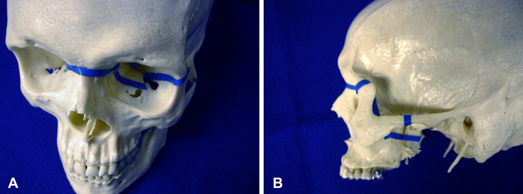
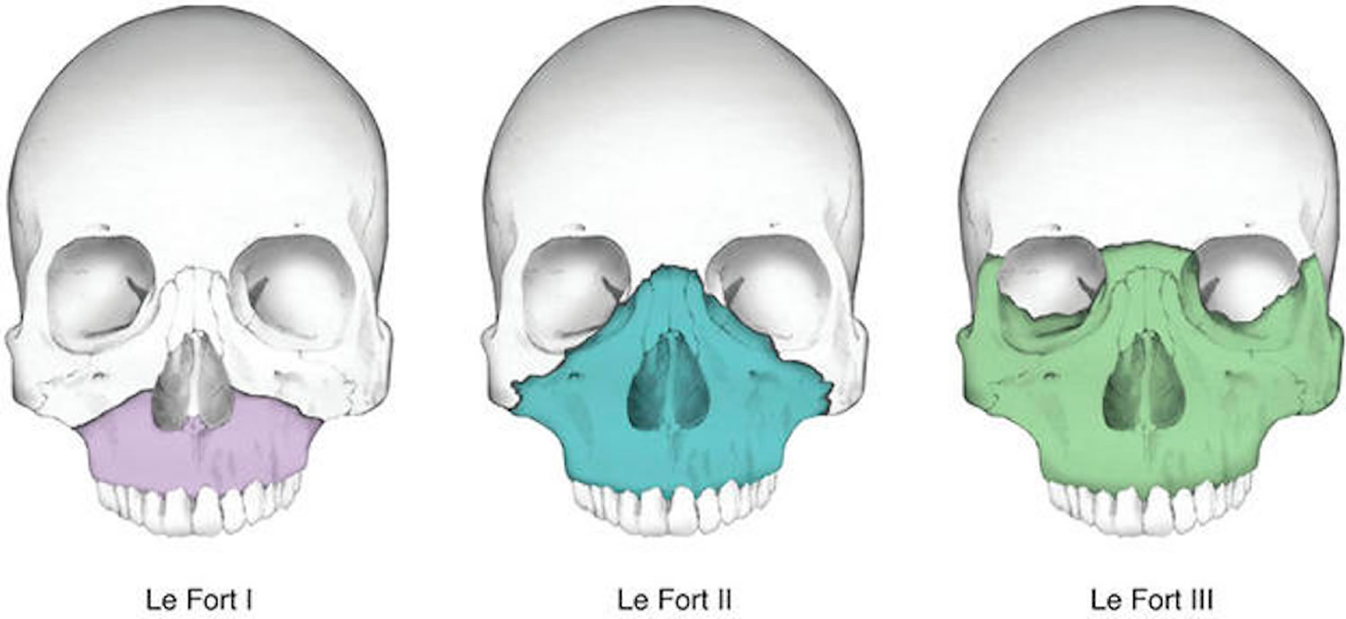
Fractures may occur in more than one plane on the same side. Do not terminate further search after identifying one Le Fort fracture. Do not rely only on clinical history and physical examination as the characteristic findings may not always be present. If one of the Le Fort fractures is suspected because of a break in its unique component, it should be confirmed by identifying the other fractures that are expected in the plane of that type of fracture.ġ. If one of these structures is intact, the corresponding type of Le Fort is excluded. Anterolateral margin of the nasal fossa (type I), To classify the type, look at the three bony structures that are unique to each type (Fig. A fracture of the pterygoid processes almost always indicates that there is at least one of the Le Fort fractures (Fig. Always look at the pterygoid processes (coronal images). Suggestive clinical signs -in a patient with a history of blunt facial trauma- include epistaxis, infraorbital ecchymosis or oedema, tenderness and separation at frontozygomatic suture, lengthening of face, depression of occular levels, enophthalmos and hooding of eyes.īest diagnostic modality of a clinically suspected midfacial injury is CT, serving the diagnosis and surgical planning.ġ. Diagnoses are made clinically and confirmed radiologically. The frequency of Le Fort types is in the order of type II > type I > type III. Le Fort and maxillary fractures accounted for 25.5% of 663 facial fractures recently reported from a level 1 trauma center. This classification includes Le Fort I, II, and III types of fractures. Type II and III injuries associated with cribriform plate disruption and CSF rhinorrheaĭifferential Diagnosis Maxillofacial TraumaĪ 3-D CT reconstruction showing a Le Fort type 1 fracture (marked by arrow).-Rene Le Fort (1901) was the first to document a tendency for specific fracture patterns of the midface to occur following direct facial trauma.LeFort I fractures are isolated to the lower face.Most commonly occur due to motor vehicle accident.Midface fracture involving the maxilla and surrounding facial structures.


 0 kommentar(er)
